
New digital radiology equipment upgrade for X-ray
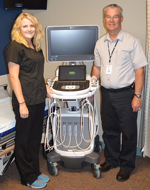
Earlier this year, the Radiology Department installed new High-Definition Ultrasound Equipment with 4D capabilities. This is especially exciting for expecting families in the community, as they will be able to view their baby in a whole new way. Pictured with the new machine is Veterans Memorial Hospital Ultrasound technicians Rachel Wager, at left, and Mike Larson, at right. 4D ultrasounds create a live video effect, like a movie, so parents can watch their baby smile, yawn or suck their thumb. The standard 2D imaging looks through the baby to show internal organs, but with the 3D and 4D scans, parents can see their baby’s skin. Submitted photo.
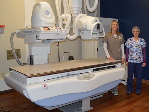
Pictured above is VMH’s fluoroscopy X-ray room which has been newly remodeled with new equipment recently installed as part of an entire digital department upgrade. Pictured left to right are X-ray Technologists Kathy Hager and Siobhan Kurth. Fluoroscopy is an imaging technique that uses X-rays to obtain real-time moving images of the interior of an object. The live images are viewed immediately for the Radiologist to evaluate and are commonly used to evaluate swallowing or gastric reflux problems. Submitted photo.
The Veterans Memorial Hospital Radiology Department has announced they have completed an upgrade to a new, fully digital radiography system which included a complete equipment upgrade and remodel of the radiology rooms.
One of the new machines that has recently been installed is a digital radiography room. Now when an X-ray exposure is taken, the image comes up instantly on the computer monitor, just like a digital camera at home. Previously, the image would be exposed onto a cassette, and run through a reader, which took about 90 seconds. The advantage of this new system is to be able to get patients done with their radiography exams quickly so they can get results faster. The new digital system not only allows for quicker exam time, but also reduces radiation dose to the patient.
Another addition to the department is a new digital fluoroscopy room. Fluoroscopy is an imaging technique that uses X-rays to obtain real-time moving images of the interior of an object. A continuous X-ray beam is passed through the body part being examined. The live images are viewed immediately for the Radiologist to evaluate. It is commonly used to evaluate swallowing or gastric reflux problems.
There is also a new digital portable X-ray machine in the department. This machine is able to go anywhere in the hospital and bring high-quality imaging right to the patient’s bedside. Since the image comes up instantly, if any repeat images need to be taken, they can be done immediately, with minimal movement of the patient, who is often in pain or has difficulty moving into different positions.
“We are ultimately making the patient experience more comfortable by efficiently delivering high quality, dose-conscious images every time,” states Amanda Leiran, Radiology Supervisor.
This new digital X-ray system produces clearer images for the radiologist to read. Since the images are computerized, the radiologist can easily adjust the brightness and contrast, or zoom in to specific areas of interest. There is always a radiologist available to read emergent exams, day or night. All digital x-rays are stored just like computer files. This is a great convenience to compare one image to another. Also, if a patient gets transferred to a larger facility, we are able to send images electronically to most area hospitals and clinics.
“We’re excited to be able to serve our patients, as well as our physicians, with better images in a more efficient and timely manner,” states Keisha Coon, X-ray Technologist. “It’s also great knowing we can reduce the amount of radiation dose we give our patients with this new equipment and technology.”
This isn’t the first time the hospital’s Radiology department has been on the cutting edge of digital technology. The computed radiology equipment at Veterans Memorial Hospital was the first of its kind in Northeast Iowa when it was installed in 2009 and has now recently been replaced.
Kelly Baxter, a new X-ray Technologist to Veterans Memorial Hospital, adds, “We are so fortunate to work in a small hospital that is willing to grow and keep up with healthcare as it continues to advance in technology every year.”
Earlier this year, the Radiology Department was the first in the area to purchase a new High-Definition Ultrasound machine with 3D and 4D capabilities. Expecting families in the community can now view their baby in a whole new way. In addition to the black and white images that are sometimes hard to visualize, there is also 3D and 4D images taken. Like regular ultrasounds, 3D and 4D ultrasounds use sound waves to create an image of the baby in the womb. What’s different is that 3D ultrasounds create a three-dimensional image of the baby, while 4D ultrasounds show moving 3D images of your baby, with time being the fourth dimension.
“Having an Ultrasound and seeing the profile of your baby is exciting as it is, but this past year our expectant families have really enjoyed seeing the baby’s entire face!” adds Supervisor Leiran.
Veterans Memorial Ultrasound services are now available five days a week. Mike Larson, Ultrasonographer at the hospital, states, “This new technology we have been offering for nearly a year now is very neat for the mothers. They really oooh and ahhh over it. The grandparents have even shared in the fun and I have heard them state, ‘This is the neatest thing since sliced bread!’ I am honored I get to see the patient reactions to this new detailed ultrasound. It’s a very neat added feature to an already very exciting time of expecting a baby.”
For more information on these digital upgrades in the Radiology Department, contact Veterans Memorial Hospital in Waukon at 563-568-3411.